Do you need MRI for low back pain? When imaging is useful
Investigating back pain starts with symptoms, nerve function and warning signs, then asks whether X-ray or MRI would guide care. Appropriate imaging can clarify a problem or support referral without making a scan necessary for every episode.
On this page
How do X-rays and MRI differ?
X-rays and magnetic resonance imaging use different physical principles and have different applications.
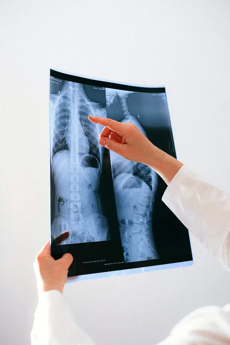
X-rays mainly assess bony structures, including spinal alignment, obvious degenerative bone changes, some fractures, spondylolisthesis and scoliosis. They involve ionising radiation and generally do not directly show discs, nerve roots or the spinal cord in detail.
MRI provides more detailed soft-tissue information, including discs, nerve roots, the spinal canal, ligaments and bone marrow. It can be important in assessing selected cases of nerve compression, infection, tumours and other specific conditions.
MRI does not use ionising radiation, but its duration, cost and suitability differ from an X-ray. Some implanted medical devices, metallic foreign bodies and other circumstances require a safety assessment before scanning.
Showing more structural detail does not mean that MRI is necessary for everyone with back pain.
Why is immediate MRI not usually needed?
NICE guidance generally advises against routine imaging for low back pain without red flags in nonspecialist settings.
One important reason is that imaging abnormalities are not always directly related to pain.
A systematic review published in 2015 analysed spinal imaging in 3,110 people without related symptoms and found disc degeneration to be common across age groups.
Its estimates included:
About 37% of asymptomatic people aged 20 had imaging evidence of disc degeneration.
About 68% of asymptomatic people aged 40 had disc degeneration.
About 88% of asymptomatic people aged 60 had disc degeneration.
These are age-related estimates pooled from studies, not an individual's personal risk.
The findings show that disc degeneration can be a common age-related imaging feature rather than a condition that necessarily needs treatment.
Even when MRI shows a disc herniation, facet degeneration or reduced disc height, imaging alone cannot establish the source of pain.
When might MRI be needed?
Although routine back pain may not require immediate investigation, the following circumstances deserve attention.
1. Suspected severe nerve compression
New difficulty passing urine, urinary retention, altered bladder or bowel control, perineal numbness or rapidly worsening neurological function may indicate an emergency such as cauda equina syndrome.
Attend an emergency department immediately. The relevant medical team can arrange urgent imaging and management.
2. Persistent or worsening nerve-root symptoms
Alongside back pain, some people develop radiating pain through the buttock, thigh and calf, sometimes with numbness or reduced strength.
If symptoms persist despite appropriate conservative treatment and surgery or another intervention is being considered, MRI may help identify the site of nerve compression and guide subsequent care.
The American College of Radiology's Appropriateness Criteria identify lumbar MRI as usually appropriate for selected patients with persistent or progressive symptoms after about six weeks of suitable management when surgery or an intervention may be needed.
Six weeks is not a mandatory waiting period for everyone. Serious or progressive neurological deficits require earlier assessment.
3. Suspected fracture, infection, tumour or other serious disease
Major trauma, fever, a history of cancer, unexplained weight loss or other concerning features may prompt appropriate imaging and further investigations.
The choice of X-ray, CT or MRI depends on the suspected condition and clinical context. MRI is not automatically the first test for everyone.
Will MRI definitely identify the cause?
Not necessarily.
MRI shows anatomical structures, whereas pain involves tissues, the nervous system and a range of physiological processes.
For example, two people may have similar disc herniations on MRI, but one has severe sciatica and the other has no symptoms.
Imaging is most useful when interpreted alongside the history and clinical examination.
When a scan does not fit the symptom distribution, treatment should not be based solely on alarming words in the report.
Should you have an X-ray first or go directly to MRI?
That depends on the clinical question identified during assessment.
An X-ray may help with some suspected fractures or alignment problems. MRI may be more appropriate for nerve compression requiring further management or selected soft-tissue conditions.
For nonspecific low back pain without red flags, neither test may be needed immediately.
Overall, the value of imaging is whether it answers an important clinical question, rather than how much detail it shows. Assessment should guide the decision, helping avoid unnecessary tests and excessive anxiety about imaging findings.
References
- Brinjikji W, et al. (2015). Systematic Literature Review of Imaging Features of Spinal Degeneration in Asymptomatic Populations. AJNR. Read study
- American College of Radiology. ACR Appropriateness Criteria: Low Back Pain. Imaging guidance
- National Institute for Health and Care Excellence. Low back pain and sciatica in over 16s: assessment and management (NG59). Clinical guideline
Further reading on low back pain
← Back to the low back pain overview
Written by: Dr Yip Siu Ho (Hong Kong Registered Chiropractor, Registration No. 441)
This article provides general health information and does not replace an individual clinical assessment.